CFAP45 deficiency causes situs abnormalities and asthenospermia by disrupting an axonemal adenine nucleotide homeostasis module
Gerard W. Dougherty, Katsutoshi Mizuno, Tabea Nöthe-Menchen, Yayoi Ikawa, Karsten Boldt, Asaf Ta-Shma, Isabella Aprea, Katsura Minegishi, Yuan-Ping Pang, Petra Pennekamp, Niki T. Loges, Johanna Raidt, Rim Hjeij, Julia Wallmeier, Huda Mussaffi, Zeev Perles, Orly Elpeleg, Franziska Rabert, Hidetaka Shiratori, Stef J. Letteboer, Nicola Horn, Samuel Young, Timo Strünker, Friederike Stumme, Claudius Werner, Heike Olbrich, Katsuyoshi Takaoka, Takahiro Ide, Wang Kyaw Twan, Luisa Biebach, Jörg Große-Onnebrink, Judith A. Klinkenbusch, Kavita Praveen, Diana C. Bracht, Inga M. Höben, Katrin Junger, Jana Gützlaff, Sandra Cindrić, Micha Aviram, Thomas Kaiser, Yasin Memari, Petras P. Dzeja, Bernd Dworniczak, Marius Ueffing, Ronald Roepman, Kerstin Bartscherer, Nicholas Katsanis, Erica E. Davis, Israel Amirav, Hiroshi Hamada & Heymut Omran, 02.11.2020
Abstract
Axonemal dynein ATPases direct ciliary and flagellar beating via adenosine triphosphate (ATP) hydrolysis. The modulatory effect of adenosine monophosphate (AMP) and adenosine diphosphate (ADP) on flagellar beating is not fully understood. Here, we describe a deficiency of cilia and flagella associated protein 45 (CFAP45) in humans and mice that presents a motile ciliopathy featuring situs inversus totalis and asthenospermia. CFAP45-deficient cilia and flagella show normal morphology and axonemal ultrastructure. Proteomic profiling links CFAP45 to an axonemal module including dynein ATPases and adenylate kinase as well as CFAP52, whose mutations cause a similar ciliopathy. CFAP45 binds AMP in vitro, consistent with structural modelling that identifies an AMP-binding interface between CFAP45 and AK8. Microtubule sliding of dyskinetic sperm from Cfap45−/− mice is rescued with the addition of either AMP or ADP with ATP, compared to ATP alone. We propose that CFAP45 supports mammalian ciliary and flagellar beating via an adenine nucleotide homeostasis module.
DOUGHERTY, Gerard W., et al. CFAP45 deficiency causes situs abnormalities and asthenospermia by disrupting an axonemal adenine nucleotide homeostasis module. Nature communications, 2020, 11. Jg., Nr. 1, S. 1-20..
Publication: https://doi.org/10.1038/s41467-020-19113-0
 Disclaimer
The publication CFAP45 deficiency causes situs abnormalities and asthenospermia by disrupting an axonemal adenine nucleotide homeostasis module by Gerard W. Dougherty, Katsutoshi Mizuno, Tabea Nöthe-Menchen, Yayoi Ikawa, Karsten Boldt, Asaf Ta-Shma, Isabella Aprea, Katsura Minegishi, Yuan-Ping Pang, Petra Pennekamp, Niki T. Loges, Johanna Raidt, Rim Hjeij, Julia Wallmeier, Huda Mussaffi, Zeev Perles, Orly Elpeleg, Franziska Rabert, Hidetaka Shiratori, Stef J. Letteboer, Nicola Horn, Samuel Young, Timo Strünker, Friederike Stumme, Claudius Werner, Heike Olbrich, Katsuyoshi Takaoka, Takahiro Ide, Wang Kyaw Twan, Luisa Biebach, Jörg Große-Onnebrink, Judith A. Klinkenbusch, Kavita Praveen, Diana C. Bracht, Inga M. Höben, Katrin Junger, Jana Gützlaff, Sandra Cindrić, Micha Aviram, Thomas Kaiser, Yasin Memari, Petras P. Dzeja, Bernd Dworniczak, Marius Ueffing, Ronald Roepman, Kerstin Bartscherer, Nicholas Katsanis, Erica E. Davis, Israel Amirav, Hiroshi Hamada & Heymut Omran is published under an open access license: http://creativecommons.org/licenses/by/4.0/. Creative Commons Attribution 4.0 International License, which permits use, sharing, adaptation, distribution and reproduction in any medium or format.
Curation by the MFGA team Relevant data sets presented in the publication have been identified. If possible, annotations (title, general information, conditions, processed tissue types and processed cell types) have been added based on information from the publication. Data tables and images that provide a good overview on the publication's findings on the data set have been extracted from the publication and/or supplement. If not stated otherwise, images are depicted with title and description exactly as in the publication. Tables have been adjusted to the MFGA table format. Conducted adjustments are explained in the detailed view of the tables. However, titles and descriptions have been adopted from the publication.
Data set 1: Loss-of-function CFAP45 mutations cause a motile ciliopathy
Exome: Whole Exome Sequencing
Species
| Species |
|---|
| Human |
| Mouse |
| Porcine |
Conditions
| Human phenotype ontology | Participants | Comment |
|---|---|---|
| HP:0012207: Reduced sperm motility | ||
| HP:control | ||
| HP:0012265: Ciliary dyskinesia | 3.0 | All three individuals presented mild chronic upper respiratory symptoms and LRA abnormalities including situs inversus totalis. |
Tissue Types
| BRENDA tissue ontology | Maturity | Description | Species | Replicates |
|---|---|---|---|---|
| BTO_0000203: respiratory system | ||||
| BTO_0002637: respiratory epithelium cell line |
Cell Types
| Cell ontology | Maturity | Description | Species | Replicates | Cells per replicate |
|---|---|---|---|---|---|
| CL_0000019: sperm |
Images
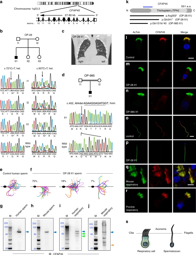
Figure. 1: CFAP45 mutations cause a motile ciliopathy.
Loss-of-function mutations in CFAP45 (a) in individuals OP-28 II1 (b) showing situs inversus totalis (note heart positioned to right rather than left side) by computerized tomography (CT) chest scan (c) and OP-985 II1 (d). Flagellar waveforms of healthy control with normospermia (e) and individual OP-28 II1 with asthenospermia (f). g–j Full-length CFAP45 (blue arrowhead, approximately 66 kilodaltons) is detectable in both human and mouse sperm and respiratory lysates; CFAP45 isoforms (green and orange arrowheads) are detectable in human and mouse respiratory lysates. Marker (M) indicates relative molecular weight (blue numerals) in kilodaltons. g–j n = 1 blot for each tissue over two experiments. k Overview of CFAP45 protein, which has a central Trichoplein domain, indicating protein change of CFAP45 mutations; blue line indicates relative epitope position of anti-CFAP45 antibody clone 3618 (amino acids 135–217). In contrast to control (l), panaxonemal CFAP45 localization (red) is undetectable in respiratory cilia from individuals OP-28 II1 (m) and OP-985 II1 (n) by IFM. In contrast to control (o), panaxonemal CFAP45 localization (red) is undetectable in sperm flagella from individual OP-28 II1 (p) by IFM. CFAP45 (red) is detectable in mouse (q) and porcine (r) respiratory cilia by IFM. Ciliary and flagellar axonemes (green) are detected using anti-acetylated α tubulin (AcTub) antibody. Merge images include Hoechst stain (blue) to indicate nuclei. White scale bars equal 10 µm. l–r n = 18 images over three experiments. s Reference cartoon for axonemes of respiratory cilia and sperm flagella.
Licensed under: http://creativecommons.org/licenses/by/4.0/
Data set 2: Proteomic profiling links axonemal CFAP45 to dynein ATPase components
Proteome: Immunohistochemistry
Species
| Species |
|---|
| Human |
| Porcine |
Cell Types
| Cell ontology | Maturity | Description | Species | Replicates | Cells per replicate |
|---|---|---|---|---|---|
| CL_0002368: respiratory epithelial cell | Porcine | Human | |||
| CL_1000497: kidney cell | HEK293 cell lysate, specifically. |
Images

Figure. 2: CFAP45 interacts with axonemal dynein complex components.
a CFAP45 immunoprecipitates from isolated porcine respiratory cells (PRC) identify 113 unique associations by LC/MS–MS including axonemal dynein complex proteins; CFAP45 was identified in all three independent experiments. b AK8 (red) is detectable in human respiratory cilia by IFM. Ciliary axonemes (green) are detected using anti-acetylated α tubulin (AcTub) antibody. Merge images include Hoechst stain (blue) to indicate nuclei. White scale bar equals 10 µm. n = 18 images over three experiments. Polyclonal anti-DNALI1 antibody immunoprecipitates myc-tagged DNALI1 (c) and FLAG-tagged CFAP45 (d), using monoclonal anti-myc and anti-FLAG antibodies by IB, respectively. Polyclonal anti-CFAP45 antibody immunoprecipitates myc-tagged CFAP45 (e) and FLAG-tagged DNALI1 (f), using monoclonal anti-myc and anti-FLAG antibodies by IB, respectively. Recombinant DNALI1 (orange arrowhead) and CFAP45 (blue arrowhead) are ~37 and 74 kilodaltons, respectively. c–f, n = 1 blot from two experiments for each IP combination. g CFAP45 directly interacts with DNALI1, as indicated by growth on LWH + 3-AT media. CFAP45 also shows binary interactions to components associated with the basal body/centrosome and intraflagellar transport (see also Supplementary Fig. 3 and Supplementary Table 3). Polyclonal anti-CFAP45 antibody immunoprecipitates native DNAH11 (h) and CFAP45 (i) from PRC lysates, using polyclonal anti-DNAH11 and anti-CFAP45 antibodies by IB. For reference, polyclonal anti-DNAH11 antibody immunoprecipitates native DNAH11 (j) but not full-length CFAP45 (k) from PRC lysates. Porcine full-length CFAP45 (blue arrowhead) and DNAH11 (green arrowhead) are ~66 and 517 kilodaltons, respectively. (h′, j′) indicate longer exposure times of (h, j) cropped above gray dashed lines. A DNAH11-immunoreactive band of ~85 kDa (red arrowhead) is detectable in both CFAP45 and DNAH11 immunoprecipitates. Species-matched (rabbit) polyclonal normal IgG is used as control. Marker (M) indicates relative molecular weight (blue numerals) in kilodaltons. h–k, n = 1 blot from two experiments for each IP combination.
Licensed under: http://creativecommons.org/licenses/by/4.0/
Data set 3: Axonemal CFAP45 abnormalities are associated with a common cause of PCD
Proteome: Immunohistochemistry
Species
| Species |
|---|
| Human |
Conditions
| Human phenotype ontology | Participants | Comment |
|---|---|---|
| HP:0012265: Ciliary dyskinesia |
Cell Types
| Cell ontology | Maturity | Description | Species | Replicates | Cells per replicate |
|---|---|---|---|---|---|
| CL_0000064: ciliated cell |
Images
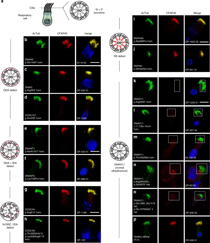
Figure. 3: Axonemal CFAP45 localization is abnormal in certain DNAH11-mutant cilia by IFM.
a Cartoon of “9 + 2” axoneme in respiratory cilia. Axonemal defects that correlate with certain PCD-causing mutations are highlighted in red. Isolated ODA defects in DNAH5- (b), DNAI2- (c), and CCDC151-mutant (d) respiratory cilia do not affect axonemal CFAP45 (red) localization. Compound ODA and IDA defects in DNAAF1- (e) and DNAAF3-mutant (f) respiratory cilia do not affect axonemal CFAP45 (red) localization. N-DRC defects lacking IDAs and with tubular disorganization in CCDC40- (g) and CCDC39-mutant (h) respiratory cilia do not affect axonemal CFAP45 (red) localization. RS defects in RSPH4A- (i) and RSPH9-mutant (j) respiratory cilia do not affect axonemal CFAP45 (red) localization. k–o Certain DNAH11-mutant respiratory cilia show abnormal ciliary CFAP45 (red) localization (rectangles). p Healthy sibling of OP-406 II2 (o) shows wild type CFAP45 localization. Ciliary axonemes (green) are detected using anti-acetylated α tubulin (AcTub) antibody. Merge images include Hoechst stain (blue) to indicate nuclei. White scale bar equals 10 µm. b–p n = 18 images from three experiments. PCD individuals with respective mutations are indicated. DNAH11 mutations are a common cause of PCD with LRA abnormalities33,34,35,36 but do not perturb the axonemal localization of ODA protein DNAH5 and IDA protein DNALI133,35. Per the study design, DNAH5 and DNALI1 are detectable by IFM in respiratory cilia from study cohort individuals (see “Methods”), suggesting that CFAP45 deficiency does not cause gross ODA and IDA abnormalities.
Licensed under: http://creativecommons.org/licenses/by/4.0/
Data set 4: CFAP45 and CFAP52 orthologs have a conserved motile ciliary function
Other: Functional Study
Species
| Species |
|---|
| Mouse |
| Multiple eukaryota |
Conditions
| Human phenotype ontology | Participants | Comment |
|---|---|---|
| HP:0012207: Reduced sperm motility | ||
| HP:control |
Cell Types
| Cell ontology | Maturity | Description | Species | Replicates | Cells per replicate |
|---|---|---|---|---|---|
| CL_0000064: ciliated cell | |||||
| CL_0000019: sperm |
Images
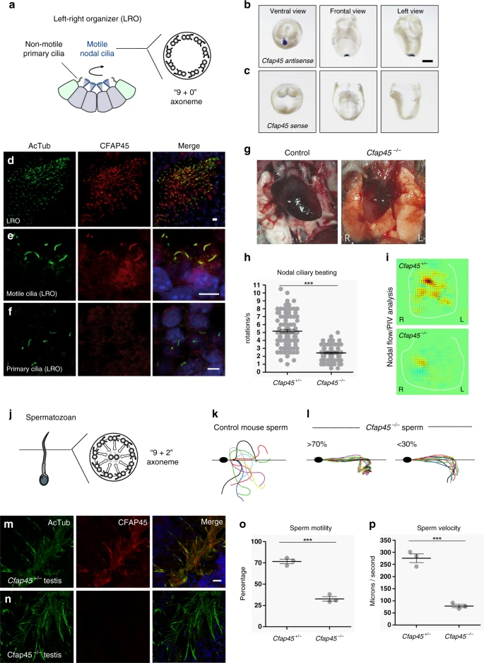
Figure. 4: Cfap45−/− mice replicate a motile ciliopathy phenotype.
a–f Motile cilia of the LRO have a “9 + 0” axoneme (a). Cfap45 is detectable in the mouse embryonic LRO by in situ hybridization (antisense probe) (b); control probe (sense) shows no hybridization (c). Black scale bar equals 100 µm. d–f CFAP45 (red) localizes to motile LRO cilia (d, e) but not non-motile (primary) cilia (f) of the LRO by IFM. g–i Compared to wild-type control, Cfap45−/− mice show LRA defects including situs inversus totalis with heart malpositioned on the right side (g). h Rotational speed of motile LRO cilia is significantly reduced in Cfap45−/− embryos (n = 74 cilia from four embryos) compared to control (n = 87 cilia from 4 embryos); data are mean ± SEM, significance assessed by two-tailed t test (***p < 0.0001). See also Supplementary Videos 3 and 4. i Particle image velocimetry (PIV) analysis shows significantly reduced fluid flow at the LRO of Cfap45−/− embryos compared to control. Yellow and red color indicate leftward flow, blue indicates rightward flow. j–p Similar to respiratory cilia, sperm flagella have a “9 + 2” axoneme (j). In contrast to control (k), Cfap45−/− sperm flagellar waveforms show reduced curvature and amplitude under conditions inducing hyperactivation (l). m, n CFAP45 (red) is detectable by IFM in testis cryosections of heterozygous littermates (m) but not Cfap45−/− males (n). o Compared to control, motility of Cfap45−/− sperm is significantly reduced (n = 195 sperm over three independent experiments for each; data are mean ± SEM; significance assessed by two-tailed t test, ***p = 0.0003). p Compared to Cfap45+/- littermates, Cfap45−/− sperm show significantly reduced curvilinear velocity (n = 99 or 70 sperm over three independent experiments for Cfap45+/− and Cfap45−/−, respectively; data are mean ± SEM; significance assessed by two-tailed t test, ***p = 0.0005). See also Supplementary Videos 5 and 6. Ciliary and flagellar axonemes (green) are detected using anti-acetylated α tubulin (AcTub) antibody. Merge images include Hoechst stain (blue) to indicate nuclei. White scale bars equal 10 µm. n = 6 images from 2 experiments (b, c, m, n); n = 4 images from one experiment in d, f.
Licensed under: http://creativecommons.org/licenses/by/4.0/
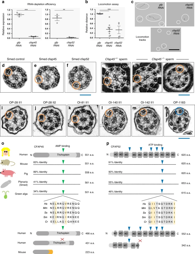
Figure. 5: CFAP45 and CFAP52 orthologs have a conserved ciliary function.
a RNAi efficiently depletes cfap45 and cfap52 in Schmidtea mediterranea (Smed) (n = 3 animals over three independent experiments for cfap45, cfap52, and gfp control); data are the means ± SEM; significance is assessed by two-tailed t-test (***p < 0.0001; **p = 0.0017). b In contrast to gfp- (n = 62 animals over eight independent experiments), the locomotion of cfap45- (n = 65 animals over eight independent experiments) and cfap52-RNAi planarians (n = 33 animals over four independent experiments) is significantly impaired in 0.6% low-melting agarose; data are the means ± SEM; significance is assessed by two-tailed t-test (***p < 0.0001). c Representative locomotion tracks (white circles) of gfp, cfap45, and cfap52-RNAi planarians; black scale bar equals 33 mm. d–n CFAP45- and CFAP52-deficient axonemes across eukaryota show normal ciliary ultrastructure. Similar to gfp control (d), TEM shows normal ciliary ultrastructure in cfap45 (e) and cfap52 (f) RNAi planarians. n = 8 images from two experiments for gfp, cfap45, and cfap52. Similar to heterozygous littermates (g), Cfap45−/− flagellar axonemes show normal ultrastructure in the midpiece (ODF outer dense fiber) and principal piece (FS fibrous sheath) regions, respectively (h). n = 8 images from two experiments for Cfap45+/− and Cfap45−/− sperm. Axonemal ultrastructure of respiratory cilia from CFAP45-deficient individual OP-28 II1 (i) is indistinguishable from healthy sibling OP-28 II2 (j) as well as CFAP52-deficient individuals OI-81 (k), OI-140 (l) and OI-142 (m). For reference, PCD individual OP-1183 (LRRC6; p.Gln188*hom.) shows a typical ciliary ultrastructure lacking both ODAs and IDAs (n). n = 6 images from 1 experiment for (i–n). Orange rings indicate detectable ODA and IDA pairs; blue ring indicates absent ODA and IDA pairs. Blue scale bars equal 100 nm. Potential AMP (green arrowhead) and ATP-binding (blue arrowhead) sites are conserved in CFAP45 (o) and CFAP52 (p) orthologs, respectively, in an isoform-specific manner. See also Fig. 1g–j and Supplementary Fig. 5g, h. Protein alignments highlight identity (gray shade) and similarity (orange shade). Red “X” indicates no predicted binding site.
Licensed under: http://creativecommons.org/licenses/by/4.0/
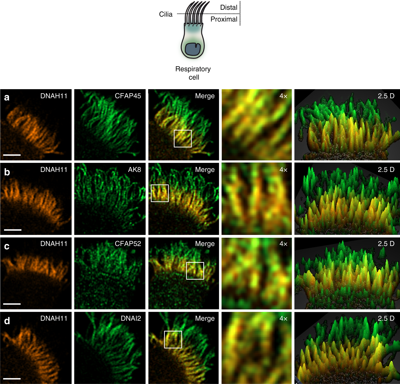
Figure. 6: CFAP45 and CFAP52 as well as AK8 colocalize with DNAH11 in the proximal region of respiratory cilia.
High-resolution Airyscan imaging of healthy control respiratory cilia shows that DNAH11 colocalizes with CFAP45 (a), AK8 (b), and CFAP52 (c) as well as the ODA component DNAI2 (d) in the proximal ciliary region. Merge panels show colocalization (yellow) in the proximal ciliary region. DNAH11 (red) is detected using monoclonal anti-DNAH11; CFAP45, CFAP52, AK8, and DNAI2 (green) are detected using respective polyclonal antibodies at a final concentration of 0.25 µg/ml. Average colocalization coefficients with reference to DNAH11 (red, Alexa Fluor 546) are as follows: CFAP45, n = 15 from two experiments (0.564, 0.663 weighted), CFAP52, n = 13 from two experiments (0.878, 0.913 weighted), AK8, n = 13 from two experiments (0.799, 0.835), DNAI2, n = 15 from two experiments (0.760, 0.820 weighted). White scale bar equals 2 µm. 4× indicates four times magnification of the white box in merge panel, which represents two square microns. Two and a half dimensional (2.5D) representations show intensity values as projections in the Z-axis (grid distance 1%).
Licensed under: http://creativecommons.org/licenses/by/4.0/
Data set 5: CFAP45 mediates adenine nucleotide homeostasis via AMP binding
Proteome: Functional Study
Species
| Species |
|---|
| Human |
Conditions
| Human phenotype ontology | Participants | Comment |
|---|---|---|
| HP:0012207: Reduced sperm motility | ||
| HP:control |
Cell Types
| Cell ontology | Maturity | Description | Species | Replicates | Cells per replicate |
|---|---|---|---|---|---|
| CL_0000019: sperm |
Images
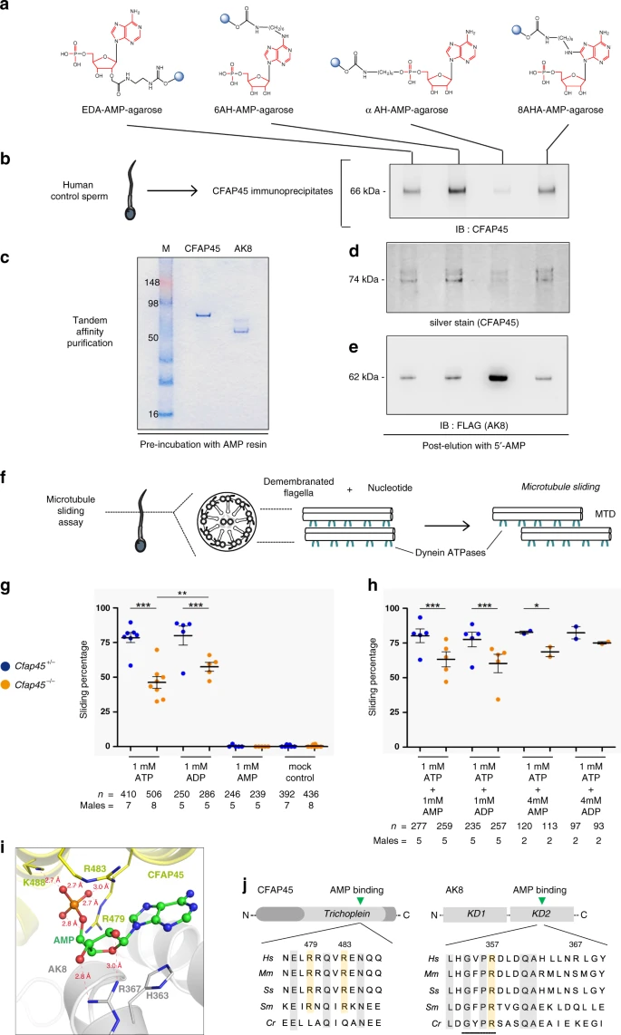
Figure. 7: CFAP45 mediates adenine nucleotide homeostasis via AMP binding.
a AMP-binding screen using AMP conjugated to agarose (blue circles) in four orientations (see also “Methods”). b CFAP45 immunoprecipitates from human sperm show affinity to AMP including 6AH-AMP-agarose following elution and IB with anti-CFAP45 antibody. c Coomasie-stained gel shows recombinant CFAP45- and AK8-enriched lysates recovered by TAP. d NTAP-CFAP45 shows affinity to AMP including 6AH-AMP-agarose by silver stain. e NTAP-AK8 shows affinity to αAH-AMP-agarose following elution and IB with monoclonal anti-FLAG antibody. f MT-sliding assay to reconstitute dynein ATPase activity. g 1 mM ATP or 1 mM ADP alone does not significantly reactivate sliding percentage of Cfap45−/− sperm compared to control (***p < 0.0001); however, 1 mM ADP significantly increases sliding percentage of Cfap45−/− sperm compared to 1 mM ATP (comparison of +/−, p = 0.3702, not significant; −/−, **p = 0.0042; data are means ± SEM; significance assessed by Fisher’s exact test); 1 mM AMP alone shows negligible (<1%) reactivation, similar to untreated control. h Combination of either 4 mM AMP (n = 113 sperm from 2 experiments, *p = 0.0432) or 4 mM ADP (n = 93 sperm over 2 experiments, p = 0.3781) with 1 mM ATP increases sliding percentage of Cfap45−/− sperm, compared to heterozygous control (data are the means ± SEM; significance supported by Fisher’s exact test); equimolar ratios of 1 mM AMP or 1 mM ADP with 1 mM ATP does not significantly reactivate Cfap45−/− sperm sliding percentage compared to control (data are means ± SEM; significance assessed by Fisher’s exact test; ***p < 0.0001). i Close-up view of AMP bound at the interface of CFAP45·AK8 complex. Carbon atoms in AMP, CFAP45, and AK8 are colored in green, yellow, and gray, respectively; nitrogen, oxygen, and phosphorus atoms are colored in blue, red, and orange, respectively. Distances shown by thin-dash lines are in Angstroms (Å). j Sequence alignments highlighting highly conserved CFAP45 residues Arg479 and Arg483- and AMP-binding site of AK8 (residues 354-357, black bar). Conserved residues highlighted in gray; arginine residues highlighted in orange. See also Supplementary Fig. 11 and “Methods”.
Licensed under: http://creativecommons.org/licenses/by/4.0/