Recessive DNAH9 Loss-of-Function Mutations Cause Laterality Defects and Subtle Respiratory Ciliary-Beating Defects
NT. Loges, Antony D, Maver A, Deardorff MA, Güleç EY, Gezdirici A, Nöthe-Menchen T, Höben IM, Jelten L, Frank D, Werner C, Tebbe J, Wu K, Goldmuntz E, Čuturilo G, Krock B, Ritter A, Hjeij R, Bakey Z, Pennekamp P, Dworniczak B, Brunner H, Peterlin B, Tanidir C, Olbrich H, Omran H, Schmidts M., 21.11.2018
Abstract
Dysfunction of motile monocilia, altering the leftward flow at the embryonic node essential for determination of left-right body asymmetry, is a major cause of laterality defects. Laterality defects are also often associated with reduced mucociliary clearance caused by defective multiple motile cilia of the airway and are responsible for destructive airway disease. Outer dynein arms (ODAs) are essential for ciliary beat generation, and human respiratory cilia contain different ODA heavy chains (HCs): the panaxonemally distributed γ-HC DNAH5, proximally located β-HC DNAH11 (defining ODA type 1), and the distally localized β-HC DNAH9 (defining ODA type 2). Here we report loss-of-function mutations in DNAH9 in five independent families causing situs abnormalities associated with subtle respiratory ciliary dysfunction. Consistent with the observed subtle respiratory phenotype, high-speed video microscopy demonstrates distally impaired ciliary bending in DNAH9 mutant respiratory cilia. DNAH9-deficient cilia also lack other ODA components such as DNAH5, DNAI1, and DNAI2 from the distal axonemal compartment, demonstrating an essential role of DNAH9 for distal axonemal assembly of ODAs type 2. Yeast two-hybrid and co-immunoprecipitation analyses indicate interaction of DNAH9 with the ODA components DNAH5 and DNAI2 as well as the ODA-docking complex component CCDC114. We further show that during ciliogenesis of respiratory cilia, first proximally located DNAH11 and then distally located DNAH9 is assembled in the axoneme. We propose that the β-HC paralogs DNAH9 and DNAH11 achieved specific functional roles for the distinct axonemal compartments during evolution with human DNAH9 function matching that of ancient β-HCs such as that of the unicellular Chlamydomonas reinhardtii.
LOGES, Niki T., et al. Recessive DNAH9 loss-of-function mutations cause laterality defects and subtle respiratory ciliary-beating defects. The American Journal of Human Genetics, 2018, 103. Jg., Nr. 6, S. 995-1008.
Publication: https://doi.org/10.1016/j.ajhg.2018.10.020
 Disclaimer
The publication Recessive DNAH9 Loss-of-Function Mutations Cause Laterality Defects and Subtle Respiratory Ciliary-Beating Defects by NT. Loges, Antony D, Maver A, Deardorff MA, Güleç EY, Gezdirici A, Nöthe-Menchen T, Höben IM, Jelten L, Frank D, Werner C, Tebbe J, Wu K, Goldmuntz E, Čuturilo G, Krock B, Ritter A, Hjeij R, Bakey Z, Pennekamp P, Dworniczak B, Brunner H, Peterlin B, Tanidir C, Olbrich H, Omran H, Schmidts M. is published under an open access no derivatives license: : https://creativecommons.org/licenses/by-nc-nd/3.0/. Permits non-commercial use of the work as published, without adaptation or alteration provided the work is fully attributed.
Curation by the MFGA team Relevant data sets presented in the publication have been identified. If possible, annotations (title, general information, conditions, processed tissue types and processed cell types) have been added based on information from the publication. Data tables and images that provide a good overview on the publication's findings on the data set have been extracted from the publication and/or supplement. If not stated otherwise, images are depicted with title and description exactly as in the publication. Tables have been adjusted to the MFGA table format. Conducted adjustments are explained in the detailed view of the tables. However, titles and descriptions have been adopted from the publication.
Data set 1: Analysis of participants with laterality defects and mild or no respiratory symptoms and PCD
Genome: Sanger Sequencing
Species
| Species |
|---|
| Human |
Conditions
| Human phenotype ontology | Participants | Comment |
|---|---|---|
| HP:other: Other | 548 | 548 individuals screened, as the total study population. |
| HP:0012265: Ciliary dyskinesia | 138 | 138 individuals were classified to have PCD with ODA defects, 255 individuals suspected to have PCD with normal ciliary structure documented by IF and/or TEM, 99 individuals suspected to have PCD without any information on the ciliary structure, 12 individuals with mild respiratory symptoms and ciliary beating defects documented by HSVM without laterality defects. |
Images
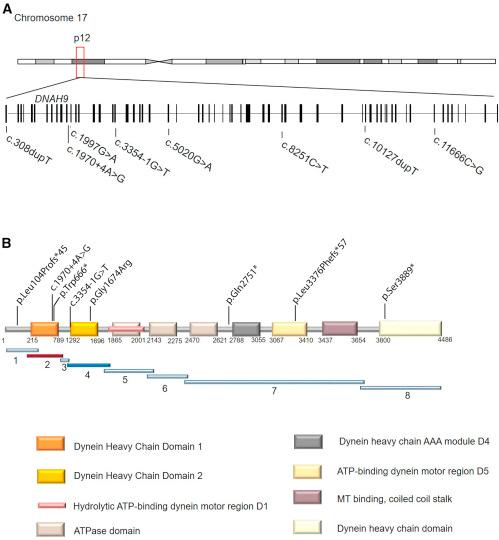
Figure 1. DNAH9 Mutations in Individuals with Laterality Defects and/or Mild Respiratory Symptoms
(A) Schematic presentation of chromosome 17 and the exon-intron structure of DNAH9. The positions of the eight mutations identified in five unrelated families are indicated.</br>(B) Predicted protein model of DNAH9 with the distinct domains and indication of DNA fragment distribution for protein-protein interaction experiments.
Licensed under: https://creativecommons.org/licenses/by-nc-nd/3.0/
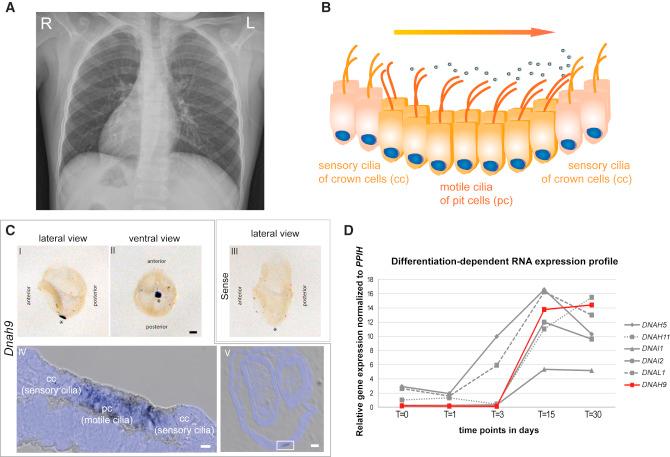
Figure 2. Dnah9 Is Essential for Determination of Left-Right Body Asymmetry and Is Expressed in Nodal Cells Carrying Motile Nodal Monocilia Important to Generate the Nodal Flow
A) The chest X-ray radiograph of MS-SI46 II1 depicts situs inversus totalis.</br>(B) Schematic of the “two cilia” nodal flow hypothesis. Motile cilia of the pit cells (pc) generate a leftward nodal fluid flow which is sensed by the sensory cilia of the crown cells (cc), which is necessary to start the signaling cascade to establish correct left-right body asymmetry.</br>(C) In situ hybridization analyses in wild-type mouse embryos show specific expression of Dnah9. Lateral (I) and ventral (II) view of a 0 somite wild-type mouse embryo demonstrate that Dnah9 expression is restricted exclusively to the left-right organizer (LRO) as indicated by the spot like dark blue signal. (III) Sense probe does not show any specific staining (I-III). Asterisks indicate the position of the LRO. Scale bars represent 100 μm. (IV) Dnah9 expression indicated by the grayish to black signal is found predominantly in the pit cells (pc) of the mouse LRO. (V) Cryosection of the embryo that previously underwent in situ hybridization using the probe directed against Dnah9. The white rectangle in V marks the region magnified in IV. (IV-V) Nuclei were counterstained with DAPI and are depicted in blue. Scale bars represent 50 μm (IV) or 5 μm (V).</br>(D) Comparison of expression profiles of genes encoding components of the outer dynein arm at five different time points of differentiation in ALI-cultured nasal epithelial cells. The RNA expression level is very low at early time points (t = 0 to t = 3) increasing heavily at later time points (t = 15) and slightly decreasing at t = 30. The differentiation-dependent expression profile of DNAH9 (in red) in ALI-cultured nasal epithelial cells resembles that of known outer dynein arm components.
Licensed under: https://creativecommons.org/licenses/by-nc-nd/3.0/
Data set 2: DNAH9 expression
Proteome: Immunohistochemistry
Species
| Species |
|---|
| Human |
Conditions
| Human phenotype ontology | Participants | Comment |
|---|---|---|
| HP:other: Other | Investigation of motile cilia function and structure in individuals with DNAH9 mutations |
Cell Types
| Cell ontology | Maturity | Description | Species | Replicates | Cells per replicate |
|---|---|---|---|---|---|
| CL_0000542: lymphocyte | Epstein-Barr-Virus-(EBV)-transformed lymphocytes (no motile cilia) (n = 2 healthy control individuals) | ||||
| CL_0000081: blood cell | (n = 3 healthy control individuals) | ||||
| nasal brushing biopsies (n = 3 healthy control individuals) | |||||
| CL_0002368: respiratory epithelial cell | respiratory cells grown on air liquid interface (ALI) culture to full differentiation (n = 2 healthy control individuals) |
Images
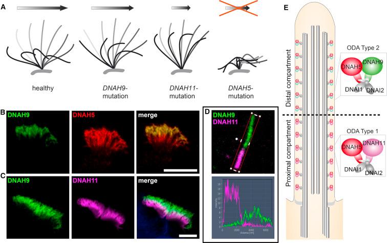
Figure 3. Schematics Depicting the Respective Aberrant Beating Patterns Caused by Recessive DNAH9, DNAH11, and DNAH5 Mutations and Localization of the Axonemal Outer Dynein Arm β-Heavy Chains (ODA HC) DNAH9 and Paralogous DNAH11 and the ODA γ-HC DNAH5 in Respiratory Cilia
(A) Schematic of representative respiratory ciliary beating defects caused by loss-of-function mutations in DNAH9, DNAH11, and DNAH5. Whereas ciliary beating in healthy individuals demonstrate effective beating (gray) and recovery (black) strokes to move mucus in the airways (arrow), DNAH9 and DNAH11 mutant cilia show reduced bending in the distal and proximal ciliary regions, respectively. DNAH5 mutant cilia are immotile with residual flickering movements.</br>(B and C) The ODA β-HC DNAH9 (green) localizes to the distal part of the ciliary axoneme and the ODA γ-HC DNAH5 (red) localizes to the whole ciliary axoneme. In contrast, the ODA β-HC DNAH11 (magenta) localizes to the proximal part of the ciliary axoneme. The overlapping front (white) results from the bending of the cilia. Scale bars represent 10 μm.</br>(D) Profiles of immunofluorescence intensity show an increase in the magenta signal in the proximal ciliary axoneme and an increase of the green signal in the distal ciliary axoneme. Both signals do not overlap indicating a distinct distal and proximal localization of β-heavy chain paralogs DNAH9 (green) and DNAH11 (magenta), respectively.</br>(E) ODA type 1 (DNAH5 and DNAH11) and ODA type 2 (DNAH5 and DNAH9) complexes define distinct compartments in respiratory cilia.
Licensed under: https://creativecommons.org/licenses/by-nc-nd/3.0/
Data set 3: Analysis of the localization of DNAH9 and effect of various DNAH mutations on ODA protein
Proteome: Immunohistochemistry
Species
| Species |
|---|
| Human |
Conditions
| Human phenotype ontology | Participants | Comment |
|---|---|---|
| HP:other: Other | 3 | Three analyzed DNAH9 mutant individuals; without a classical PCD phenotype. |
Tissue Types
| BRENDA tissue ontology | Maturity | Description | Species | Replicates |
|---|---|---|---|---|
| BTO_0000203: respiratory system |
Cell Types
| Cell ontology | Maturity | Description | Species | Replicates | Cells per replicate |
|---|---|---|---|---|---|
| CL_0002368: respiratory epithelial cell | Respiratory ciliary cells |
Images
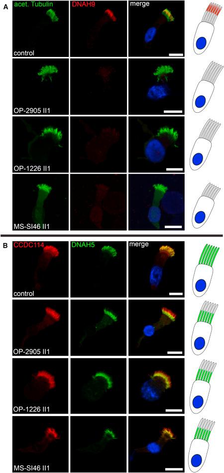
Figure 4. DNAH9 Mutant Cilia Disrupt ODA Type 2 Complexes in the Distal Compartment of Respiratory Cilia
(A) In contrast to healthy control subjects, DNAH9 (red) is absent or severely reduced in the distal compartment of DNAH9 mutant cilia. Ciliary axonemes are counterstained with anti-acetylated tubulin (green). Schematic (right side) highlights loss of DNAH9 (red) in the distal compartment of respiratory cilia.</br>(B) In contrast to healthy control subjects, DNAH5 (green) is absent or severely reduced in the distal compartment of DNAH9 mutant cilia. DNAH5 retains localization in the proximal compartment of DNAH9 mutant cilia. The ODA docking complex protein CCDC114 (red) localizes along the entire ciliary axoneme in both control and DNAH9 mutant cilia. Schematic (right side) highlights loss of DNAH5 (green) in the distal compartment of respiratory cilia. Nuclei are stained with Hoechst33342 (blue). Scale bars represent 10 μm. For each individual, 15–20 cells were analyzed from two independent IF experiments.
Licensed under: https://creativecommons.org/licenses/by-nc-nd/3.0/
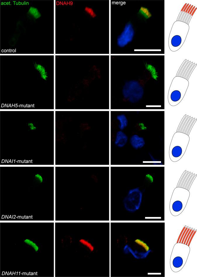
Figure 5. Mutations in DNAH5, DNAI1, DNAI2, and DNAH11 Affect Axonemal Localization of DNAH9
Respiratory cilia double-labeled with antibodies directed against acetylated tubulin (green) and DNAH9 (red) show colocalization of DNAH9 with acetylated tubulin at the distal part of the cilia from an unaffected control (yellow). In contrast, DNAH9 is absent or severely reduced in DNAH5, DNAI1, and DNAI2 mutant ciliary axonemes. Interestingly, in DNAH11 mutant cilia the distal localization of DNAH9 shifts from distal to a panaxonemal localization. Nuclei were stained with Hoechst33342 (blue). Scale bars represent 10 μm. For each individual, 15–20 cells were analyzed.
Licensed under: https://creativecommons.org/licenses/by-nc-nd/3.0/
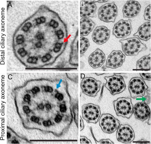
Figure 6. Distal Ciliary Axonemes of DNAH9-Deficient Respiratory Cells Display Subtle Ultrastructural Defects of the Outer Dynein Arms (ODAs)
(A and B) Loss-of-function mutations in DNAH9 cause distal absence of ODAs in the transmission-electron microscopy but do not affect the ODA docking complex (DC) (red arrow in A).</br>(C and D) Interestingly, ODAs in the proximal part of the ciliary axonemes (blue arrow in C) are not affected by DNAH9 deficiency. The green arrow in (D) marks microvilli extending from the apical side of the cell.</br>Scale bars represent 200 nm. A total of 153 ciliary cross-sections were analyzed.
Licensed under: https://creativecommons.org/licenses/by-nc-nd/3.0/
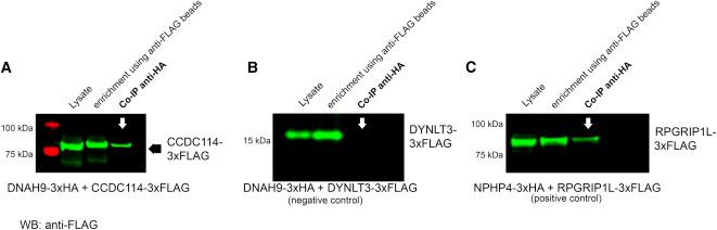
Figure 7. DNAH9 DHC Domain 1 Interacts with CCDC114
Co-immunoprecipitation (co-IP) confirmed the interaction between DNAH9 fragment 2 and CCDC114. HEK293T cells were transfected with DNAH9-fragment-2-3xHA and CCDC114-3xFLAG (A), DNAH9 fragment-2-3xHA, and DYNLT3-3xFLAG (B, negative control). NPHP4-3xHA and RPGRIP1L-C-term-3xFLAG were used as positive control (C). Co-IP was performed with the anti-HA antibody while anti-FLAG beads were used for enrichment. Western blot (WB) was performed using an antibody directed against the FLAG tag. DNAH9-fragment-2 was able to precipitate full-length CCDC114 (A) with protein amounts detected in the co-IP comparable to what we observe for the positive control (C) while no interaction between DNAH9-fragment-2 and DYNLT3 was observed (negative control) (B). Lysate shows protein content before pull down, enrichment shows pulled down prey protein.
Licensed under: https://creativecommons.org/licenses/by-nc-nd/3.0/