Dissecting the spermatogonial stem cell niche using spatial transcriptomics
Rajachandran S, Zhang X, Cao Q, Caldeira-Brant AL, Zhang X, Song Y, Evans M, Bukulmez O, Grow EJ, Nagano M, Orwig KE, Chen H., 25.07.2023
Abstract
Spermatogonial stem cells (SSCs) in the testis support the lifelong production of sperm. SSCs reside within specialized microenvironments called "niches," which are essential for SSC self-renewal and differentiation. However, our understanding of the molecular and cellular interactions between SSCs and niches remains incomplete. Here, we combine spatial transcriptomics, computational analyses, and functional assays to systematically dissect the molecular, cellular, and spatial composition of SSC niches. This allows us to spatially map the ligand-receptor (LR) interaction landscape in both mouse and human testes. Our data demonstrate that pleiotrophin regulates mouse SSC functions through syndecan receptors. We also identify ephrin-A1 as a potential niche factor that influences human SSC functions. Furthermore, we show that the spatial re-distribution of inflammation-related LR interactions underlies diabetes-induced testicular injury. Together, our study demonstrates a systems approach to dissect the complex organization of the stem cell microenvironment in health and disease.
Rajachandran S, Zhang X, Cao Q, Caldeira-Brant AL, Zhang X, Song Y, Evans M, Bukulmez O, Grow EJ, Nagano M, Orwig KE, Chen H. Dissecting the spermatogonial stem cell niche using spatial transcriptomics. Cell Rep. 2023 Jul 25;42(7):112737. doi: 10.1016/j.celrep.2023.112737. Epub 2023 Jul 1. PMID: 37393620.
Publication: https://doi.org/10.1016/j.celrep.2023.112737
 Disclaimer
The publication Dissecting the spermatogonial stem cell niche using spatial transcriptomics by Rajachandran S, Zhang X, Cao Q, Caldeira-Brant AL, Zhang X, Song Y, Evans M, Bukulmez O, Grow EJ, Nagano M, Orwig KE, Chen H. is published under an open access license: https://creativecommons.org/licenses/by/4.0/. Permits non-commercial re-use, distribution, and reproduction in any medium, provided the original work is properly cited.
Curation by the MFGA team Relevant data sets presented in the publication have been identified. If possible, annotations (title, general information, conditions, processed tissue types and processed cell types) have been added based on information from the publication. Data tables and images that provide a good overview on the publication's findings on the data set have been extracted from the publication and/or supplement. If not stated otherwise, images are depicted with title and description exactly as in the publication. Tables have been adjusted to the MFGA table format. Conducted adjustments are explained in the detailed view of the tables. However, titles and descriptions have been adopted from the publication.
Data set 1: A spatially resolved LR interaction map of the mouse testis
Transcriptome: Slide-seq
Species
| Species |
|---|
| Mouse |
Tissue Types
| BRENDA tissue ontology | Maturity | Description | Species | Replicates |
|---|---|---|---|---|
| BTO_0001363: testis | adult (7–18 weeks old) | A typically paired male reproductive gland that produces sperm and that in most mammals is contained within the scrotum at sexual maturity. | Mouse |
Cell Types
| Cell ontology | Maturity | Description | Species | Replicates | Cells per replicate |
|---|---|---|---|---|---|
| CL_4030037: late spermatid | elongated spermatids | Mouse | |||
| CL_4030036: early spermatid | round spermatids | Mouse | |||
| CL_0000017: spermatocyte | A male germ cell that develops from spermatogonia. The euploid primary spermatocytes undergo meiosis and give rise to the haploid secondary spermatocytes which in turn give rise to spermatids. | Mouse | |||
| CL_0000020: spermatogonium | An euploid male germ cell of an early stage of spermatogenesis. | Mouse | |||
| CL_0000115: endothelial cell | An endothelial cell comprises the outermost layer or lining of anatomical structures and can be squamous or cuboidal. In mammals, endothelial cell has vimentin filaments and is derived from the mesoderm. | Mouse | |||
| CL_0000178: Leydig cell | A Leydig cell is a testosterone-secreting cell in the interstitial area, between the seminiferous tubules, in the testis. | Mouse | |||
| CL_0000216: Sertoli cell | A supporting cell projecting inward from the basement membrane of seminiferous tubules. They surround and nourish the developing male germ cells and secrete androgen binding protein. Their tight junctions with the spermatogonia and spermatocytes provide a blood-testis barrier. | Mouse |
Images
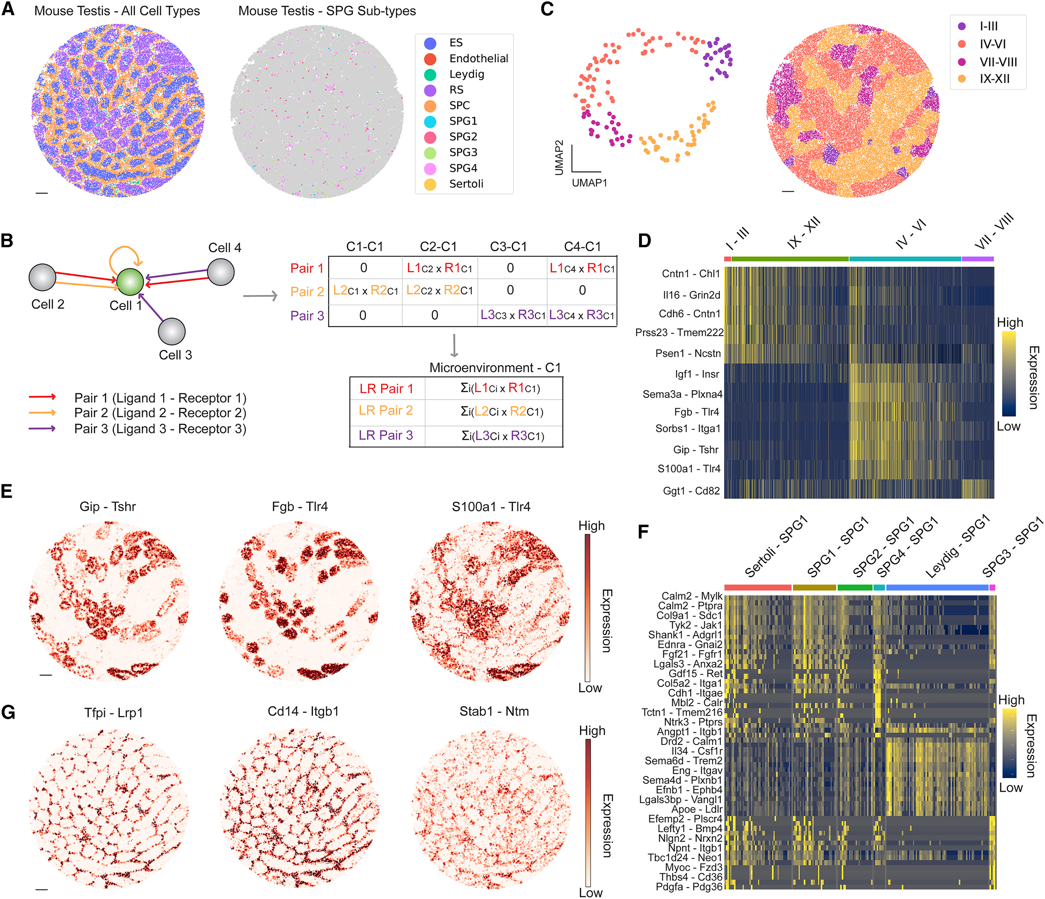
Figure 1: A spatially resolved ligand-receptor (LR) interaction map of the mouse testis
(A) Spatial mapping of major testicular cell types (left) and transcriptional states of spermatogonia (right) using a mouse testis Slide-seq dataset. ES, elongating/elongated spermatid; RS, round spermatid; SPC, spermatocyte; SPG, spermatogonium. Scale bar, 150 μm. (B) Schematic of the method to calculate spatially resolved LR interactions between a set of cells. Cell-cell interactions are calculated by multiplying ligand expression on the sending cell with receptor expression on the neighboring receiving cell for each LR pair. (C) Left: uniform manifold approximation and projection (UMAP) of seminiferous tubules in the transcriptome space colored by the stages of the cycle of the seminiferous epithelium. Right: spatial mapping of the stages of the cycle of the seminiferous epithelium. Scale bar, 150 μm. (D) Differentially expressed LR pairs across the stages of the seminiferous epithelium cycle. (E) Spatial expression patterns of selective LR pairs enriched in stages IV–VI of the seminiferous epithelium cycle. Scale bar, 160 μm. (F) Differentially expressed LR pairs among cell type-SPG1 pairs. The heatmap shows 60 representative LR pairs. Every other row of the heatmap is labeled because of space limitations. The full list of LR pairs is provided in Table S1. (G) Spatial expression patterns of selective LR pairs enriched in the Leydig cell-SPG1 pair. Scale bar, 160 μm.
Licensed under: https://creativecommons.org/licenses/by/4.0/
Data set 2: PTN-mediated signaling regulates the functions of mouse SSCs
Other: Immunohistochemistry
Species
| Species |
|---|
| Mouse |
Tissue Types
| BRENDA tissue ontology | Maturity | Description | Species | Replicates |
|---|---|---|---|---|
| BTO_0001363: testis | adult (7–18 weeks old) | A typically paired male reproductive gland that produces sperm and that in most mammals is contained within the scrotum at sexual maturity. | Mouse |
Cell Types
| Cell ontology | Maturity | Description | Species | Replicates | Cells per replicate |
|---|---|---|---|---|---|
| CL_0000020: spermatogonium | An euploid male germ cell of an early stage of spermatogenesis. | Mouse | |||
| PLANA_0000219: spermatogonial stem cell | SSC; Gh4+, nanos+ cells residing at the testis periphery that self-renew and give rise to differentiating daughters that will ultimately produce haploid sperm. | Mouse |
Images
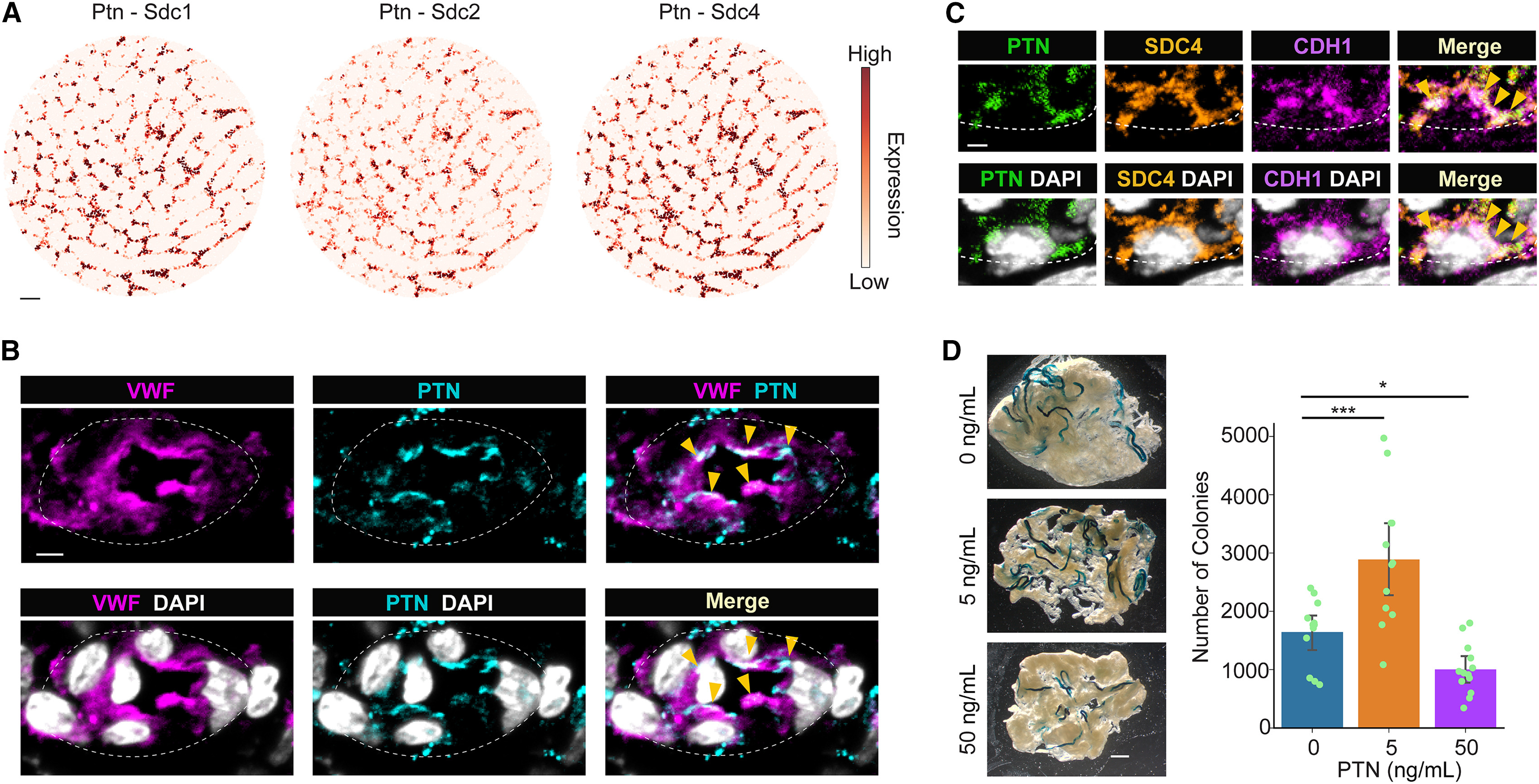
Figure 2: PTN-mediated signaling regulates the functions of mouse SSCs
(A) Spatial expression patterns of Ptn-Sdc pairs revealed by the mouse testis Slide-seq data. Scale bar, 160 μm. (B) Endothelial cell marker protein VWF co-localizes with PTN protein (yellow arrowheads). The white dashed line outlines the blood vessel. Scale bar, 3 μm. (C) Co-localization of PTN and SDC4 protein in a CDH1+ undifferentiated spermatogonium (yellow arrowheads). The white dashed line outlines the basement membrane of the seminiferous tubule. Scale bar, 2 μm. (D) Cultured mouse SSCs treated with PTN protein at 0, 5, or 50 ng/mL were transplanted into busulfan-treated recipients (n = 12, n = 12, and n = 13 recipient testes for the 0, 5, and 50 ng/mL group, respectively). Colony numbers in recipient testes were determined two months later using the LacZ staining. Colony counts were normalized as “per culture well” to make fair comparisons across groups. Data are represented as mean ± SD. One-way ANOVA followed by Fisher’s least significant difference test; ∗p < 0.05 and ∗∗∗p < 0.001. Representative images of the LacZ-stained recipient testes for each group are shown. Scale bar, 1 mm.
Licensed under: https://creativecommons.org/licenses/by/4.0/
Data set 3: Global cell-cell communications in the mouse testis
Transcriptome: Slide-seq
Species
| Species |
|---|
| Mouse |
Tissue Types
| BRENDA tissue ontology | Maturity | Description | Species | Replicates |
|---|---|---|---|---|
| BTO_0001363: testis | adult (7–18 weeks old) | A typically paired male reproductive gland that produces sperm and that in most mammals is contained within the scrotum at sexual maturity. | Mouse |
Cell Types
| Cell ontology | Maturity | Description | Species | Replicates | Cells per replicate |
|---|---|---|---|---|---|
| CL_0000018: spermatid | A male germ cell that develops from the haploid secondary spermatocytes. Without further division, spermatids undergo structural changes and give rise to spermatozoa. | Mouse | |||
| CL_0000017: spermatocyte | A male germ cell that develops from spermatogonia. The euploid primary spermatocytes undergo meiosis and give rise to the haploid secondary spermatocytes which in turn give rise to spermatids. | Mouse | |||
| CL_0000020: spermatogonium | An euploid male germ cell of an early stage of spermatogenesis. | Mouse | |||
| CL_0000115: epididymal spermatozoa | An endothelial cell comprises the outermost layer or lining of anatomical structures and can be squamous or cuboidal. In mammals, endothelial cell has vimentin filaments and is derived from the mesoderm. | Mouse | |||
| CL_0000178: Leydig cell | A Leydig cell is a testosterone-secreting cell in the interstitial area, between the seminiferous tubules, in the testis. | Mouse | |||
| CL_0000216: Sertoli cell | A supporting cell projecting inward from the basement membrane of seminiferous tubules. They surround and nourish the developing male germ cells and secrete androgen binding protein. Their tight junctions with the spermatogonia and spermatocytes provide a blood-testis barrier. | Mouse |
Images
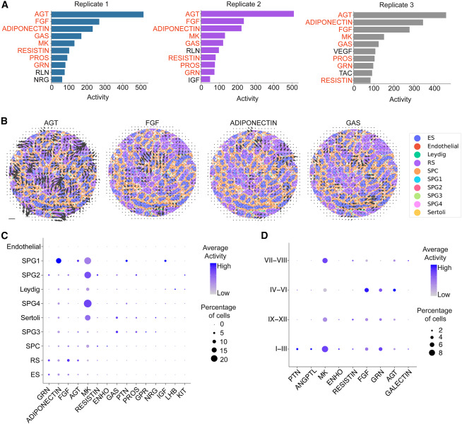
Figure 3: Global signaling activities in the mouse testis
(A) The top 10 signaling pathways with the highest activities across 3 replicates of mouse testis Slide-seq datasets. Pathways in red are shared across replicates. (B) The signaling directions of four signaling pathways with high activities. The size of the arrow is proportional to the strength of the signaling activity. Scale bar, 200 μm. (C) Dot plot showing the differential signaling activities received by each mouse testicular cell type. (D) Dot plot showing the differential signaling activities received by cells in each stage of the cycle of the seminiferous epithelium.
Licensed under: https://creativecommons.org/licenses/by/4.0/
Data set 4: A spatially resolved LR interaction map of the human testis
Transcriptome: Slide-seq
Species
| Species |
|---|
| Human |
Tissue Types
| BRENDA tissue ontology | Maturity | Description | Species | Replicates |
|---|---|---|---|---|
| BTO_0001363: testis | adult | A typically paired male reproductive gland that produces sperm and that in most mammals is contained within the scrotum at sexual maturity. | Human | 4 |
Cell Types
| Cell ontology | Maturity | Description | Species | Replicates | Cells per replicate |
|---|---|---|---|---|---|
| CL_4030037: late spermatid | adult | elongated spermatids | Human | ||
| CL_4030036: early spermatid | adult | round spermatids | Human | ||
| CL_0002481: peritubular myoid cell | adult | The flattened smooth myoepithelial cells of mesodermal origin that lie just outside the basal lamina of the seminiferous tubule. | Human | ||
| CL_0000235: macrophage | adult | A mononuclear phagocyte present in variety of tissues, typically differentiated from monocytes, capable of phagocytosing a variety of extracellular particulate material, including immune complexes, microorganisms, and dead cells. | Human | ||
| CL_0000017: spermatocyte | adult | A male germ cell that develops from spermatogonia. The euploid primary spermatocytes undergo meiosis and give rise to the haploid secondary spermatocytes which in turn give rise to spermatids. | Human | ||
| CL_0000020: spermatogonium | adult | An euploid male germ cell of an early stage of spermatogenesis. | Human | ||
| CL_0000115: endothelial cell | adult | An endothelial cell comprises the outermost layer or lining of anatomical structures and can be squamous or cuboidal. In mammals, endothelial cell has vimentin filaments and is derived from the mesoderm. | Human | ||
| CL_0000178: Leydig cell | adult | A Leydig cell is a testosterone-secreting cell in the interstitial area, between the seminiferous tubules, in the testis. | Human | ||
| CL_0000216: Sertoli cell | adult | A supporting cell projecting inward from the basement membrane of seminiferous tubules. They surround and nourish the developing male germ cells and secrete androgen binding protein. Their tight junctions with the spermatogonia and spermatocytes provide a blood-testis barrier. | Mouse |
Images
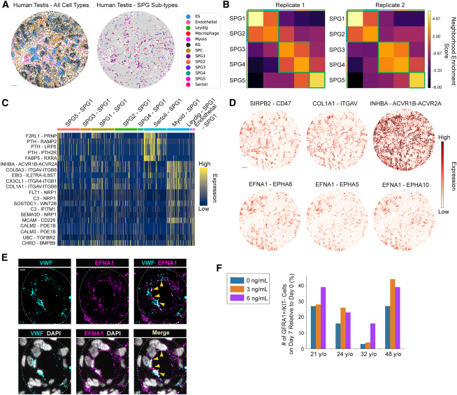
Figure 4: A spatially resolved LR interaction map of the human testis
(A) Spatial mapping of major testicular cell types (left) and transcriptional states of spermatogonia (right) in a human testis sample. ES, elongating/elongated spermatid; RS, round spermatid; SPC, spermatocyte; SPG, spermatogonium. Scale bar, 300 μm. (B) The spatial relationship between the five SPG transcriptional subtypes as quantified by the neighborhood enrichment score in two independent human Slide-seq datasets. (C) Differentially expressed LR pairs among SPG1-neighboring cell type pairs. (D) Spatial expression patterns of selective LR pairs enriched in myoid cell-SPG1 (upper panel) and endothelial cell-SPG1 (lower panel) pairs. Scale bar, 320 μm. (E) Protein expression of VWF and EFNA1 in a human testicular sample. The white dashed line outlines the blood vessel. Yellow arrowheads denote protein co-localization. Scale bar, 8 μm. (F) Bar graph showing the number of GFRA1+; KIT− human SPG on day 7 normalized against that from day 0. Cells were treated with recombinant human ENFA protein at 0, 3, and 6 ng/mL, respectively. Data from 4 patient samples are shown.
Licensed under: https://creativecommons.org/licenses/by/4.0/