Mutations in PIH1D3 Cause X-Linked Primary Ciliary Dyskinesia with Outer and Inner Dynein Arm Defects
Tamara Paff, Niki T.Loges, Isabella Aprea, Kaman Wu, Zeineb Bakey, Eric G. Haarman, Johannes M.A. Daniels, Erik A. Sistermans, Natalija Bogunovic, Gerard W. Dougherty, Inga M.Höben, Jörg Große-Onnebrink, Anja Matter, Heike Olbrich, Claudius Werner, Gerard Pals, Miriam Schmidts, Heymut Omran, Dimitra Micha, 29.12.2016
Abstract
Defects in motile cilia and sperm flagella cause primary ciliary dyskinesia (PCD), characterized by chronic airway disease, infertility, and left-right body axis disturbance. Here we report maternally inherited and de novo mutations in PIH1D3 in four men affected with PCD. PIH1D3 is located on the X chromosome and is involved in the preassembly of both outer (ODA) and inner (IDA) dynein arms of cilia and sperm flagella. Loss-of-function mutations in PIH1D3 lead to absent ODAs and reduced to absent IDAs, causing ciliary and flagellar immotility. Further, PIH1D3 interacts and co-precipitates with cytoplasmic ODA/IDA assembly factors DNAAF2 and DNAAF4. This result has clinical and genetic counseling implications for genetically unsolved male case subjects with a classic PCD phenotype that lack additional phenotypes such as intellectual disability or retinitis pigmentosa.
PAFF, Tamara, et al. Mutations in PIH1D3 cause X-linked primary ciliary dyskinesia with outer and inner dynein arm defects. The American Journal of Human Genetics, 2017, 100. Jg., Nr. 1, S. 160-168.
Publication: https://doi.org/10.1016/j.ajhg.2016.11.019
 Disclaimer
The publication Mutations in PIH1D3 Cause X-Linked Primary Ciliary Dyskinesia with Outer and Inner Dynein Arm Defects by Tamara Paff, Niki T.Loges, Isabella Aprea, Kaman Wu, Zeineb Bakey, Eric G. Haarman, Johannes M.A. Daniels, Erik A. Sistermans, Natalija Bogunovic, Gerard W. Dougherty, Inga M.Höben, Jörg Große-Onnebrink, Anja Matter, Heike Olbrich, Claudius Werner, Gerard Pals, Miriam Schmidts, Heymut Omran, Dimitra Micha is published under an open access no derivatives license: : https://creativecommons.org/licenses/by-nc-nd/4.0/. Granted rights: share — copy and redistribute the material in any medium or format. No alterations allowed. Thus, tables can not be shown in MFGA format.
Curation by the MFGA team Relevant data sets presented in the publication have been identified. If possible, annotations (title, general information, conditions, processed tissue types and processed cell types) have been added based on information from the publication. Data tables and images that provide a good overview on the publication's findings on the data set have been extracted from the publication and/or supplement. If not stated otherwise, images are depicted with title and description exactly as in the publication. Tables have been adjusted to the MFGA table format. Conducted adjustments are explained in the detailed view of the tables. However, titles and descriptions have been adopted from the publication.
Data set 1:
Other: Functional Study
Species
| Species |
|---|
| Human |
Conditions
| Human phenotype ontology | Participants | Comment |
|---|---|---|
| HP:0012265: Ciliary dyskinesia |
Cell Types
| Cell ontology | Maturity | Description | Species | Replicates | Cells per replicate |
|---|---|---|---|---|---|
| CL_0000066: epithelial cell | Adult | Respiratory epithelial cells | Human | ||
| CL_0000019: sperm | Adult | Human |
Images
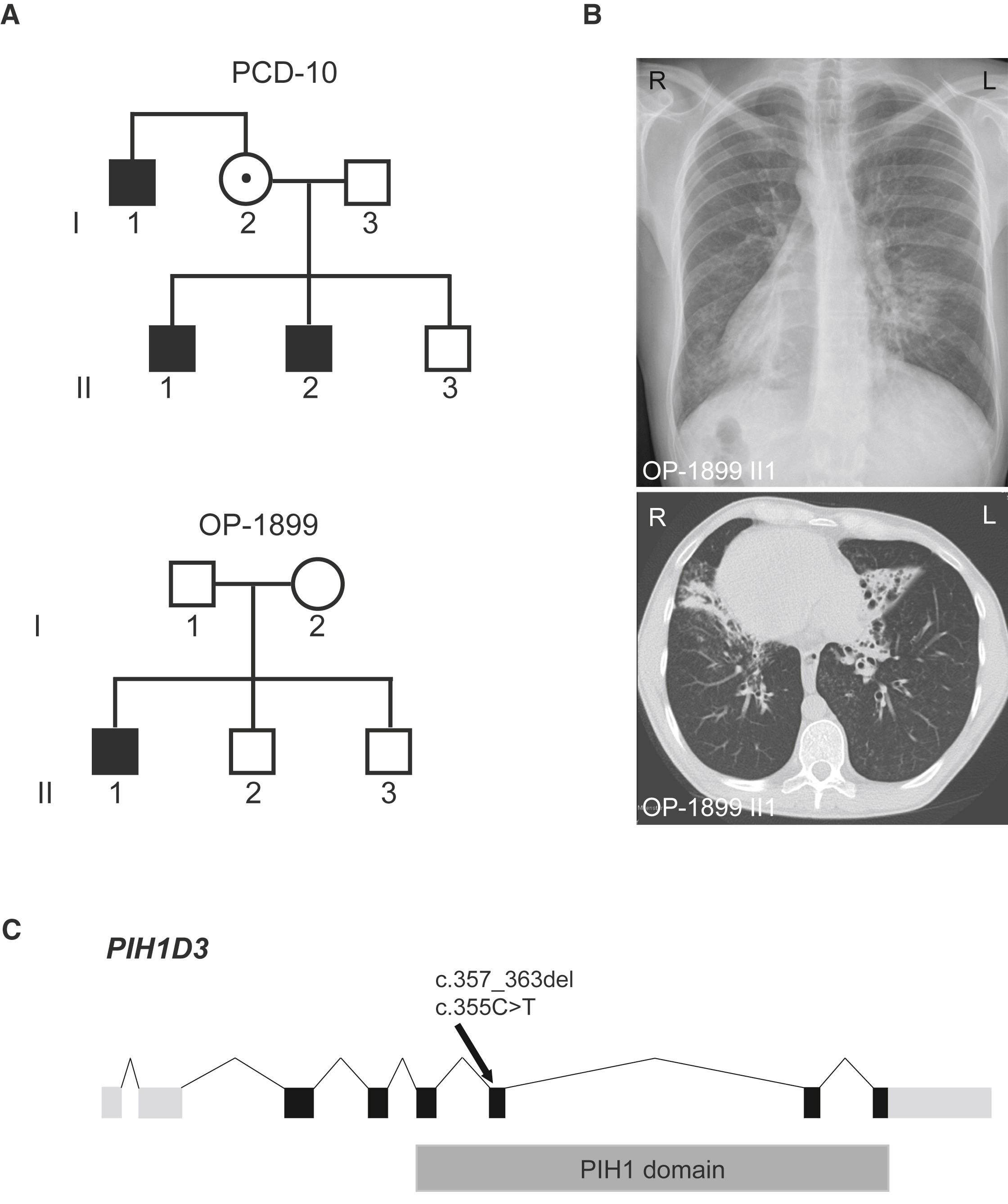
Figure 1: Results of the Mutational Analyses in PIH1D3, Pedigrees of the PCD-Affected Families PCD-10 and OP-1899, and Clinical Features of OP-1899 II1
(A) Hemizygous loss-of-function mutations in PIH1D3 located on the X chromosome were identified in two families. Pedigree of PCD-affected families PCD-10 and OP-1899. PCD-affected siblings are shaded black and the unaffected sibling is shaded white.</br>(B) The chest X-ray radiograph as well as two computed tomography scans show situs inversus totalis, chronic airway disease with bronchiectasis in the middle lobe, and mucus plugging in OP-1899 II1.</br>(C) Exon-intron structure of PIH1D3. The exon-intron structure of PIH1D3 with untranslated (light gray) and translated (black) regions and the PIH1 domain (dark gray).
Licensed under: https://creativecommons.org/licenses/by-nc-nd/4.0/
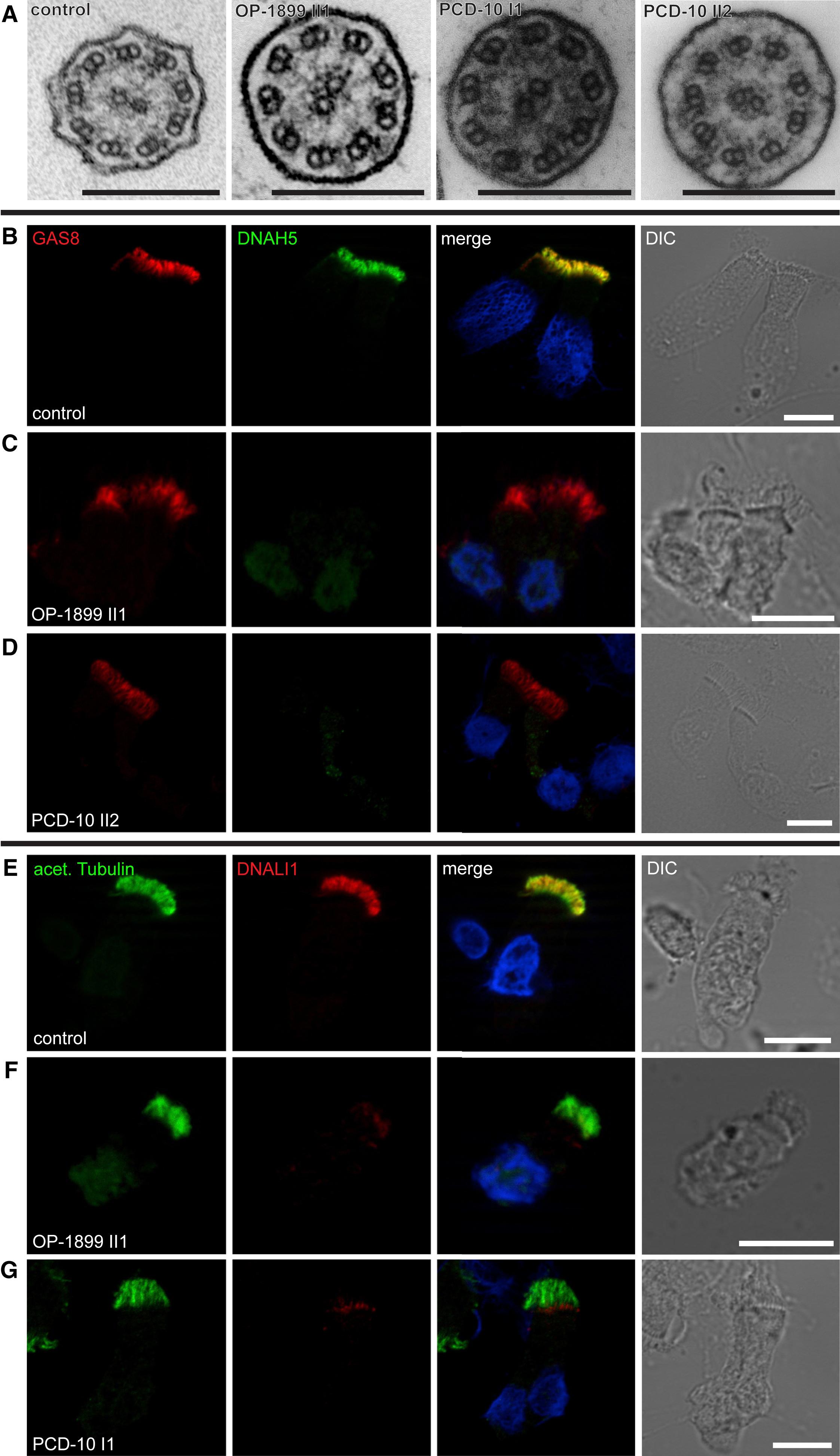
Figure 2: Loss-of-Function PIH1D3 Mutations Result in Absence of Outer and Inner Dynein Arms in Respiratory Epithelial Cells
(A) The analysis of transmission electron microscopy cross-sections from respiratory cilia from OP-1899 II1, PCD-10 I1, and PCD-10 II2 showed a normal 9+2 architecture with absence of outer dynein and inner dynein arms. Scale bars represent 200 nm.</br>(B–D) Respiratory epithelial cells from control (B) and PCD-affected individuals OP-1899 II1 (C) and PCD-10 II2 (D) were double-labeled with antibodies directed against outer dynein arm heavy chain DNAH5 (green) and the N-DRC component GAS8 (red). Both proteins colocalize along the cilia in cells from the unaffected controls (B, yellow). In contrast, in mutant cells, DNAH5 was absent from or severely reduced in ciliary axonemes (C and D). The green staining in the nuclei observed in (C) is most probably background.</br>(E–G) Cells were double-labeled with antibodies directed against acetylated tubulin (green) and the inner dynein arm light chain DNALI1 (red). In unaffected control cilia, both proteins colocalize along the ciliary axonemes (E, yellow). In contrast, in cells of individuals carrying PIH1D3 mutations, DNALI1 was absent from or severely reduced in the ciliary axonemes (F and G). The red staining at the ciliary base is an artifact caused by the polyclonal rabbit antibody. Nuclei were stained with Hoechst33342 (blue). Scale bars represent 10 μm.
Licensed under: https://creativecommons.org/licenses/by-nc-nd/4.0/
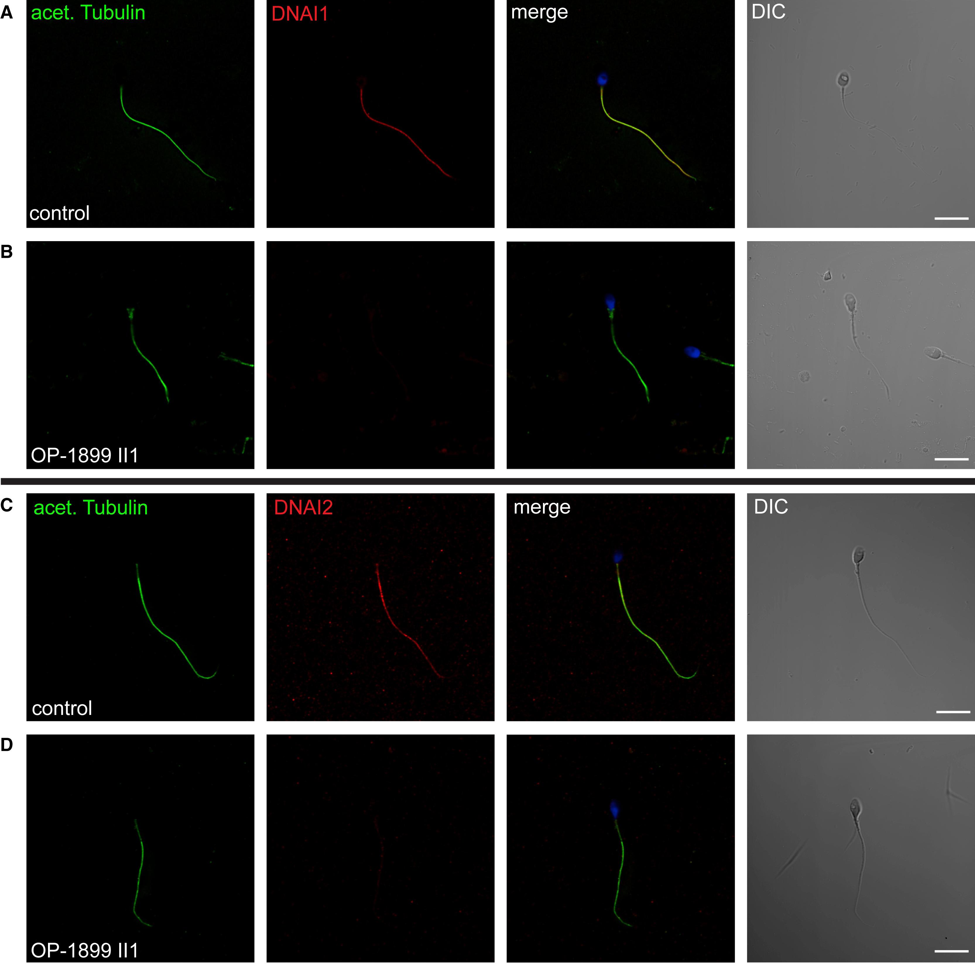
Figure 3: PIH1D3 Mutant Sperm Flagella Are Deficient for the Outer Dynein Arm Intermediate Chains DNAI1 and DNAI2
Sperms were double-labeled with antibodies directed against acetylated tubulin (green) and DNAI1 or DNAI2 (both in red). DNAI1 and DNAI2 colocalize with acetylated tubulin along the flagellum in sperms from unaffected controls (yellow) (A and C). In contrast, in PIH1D3 mutant sperm tails, DNAI1 (B) and DNAI2 (D) were absent from flagellar axonemes. Nuclei were stained with Hoechst33342 (blue). Scale bars represent 10 μm.
Licensed under: https://creativecommons.org/licenses/by-nc-nd/4.0/
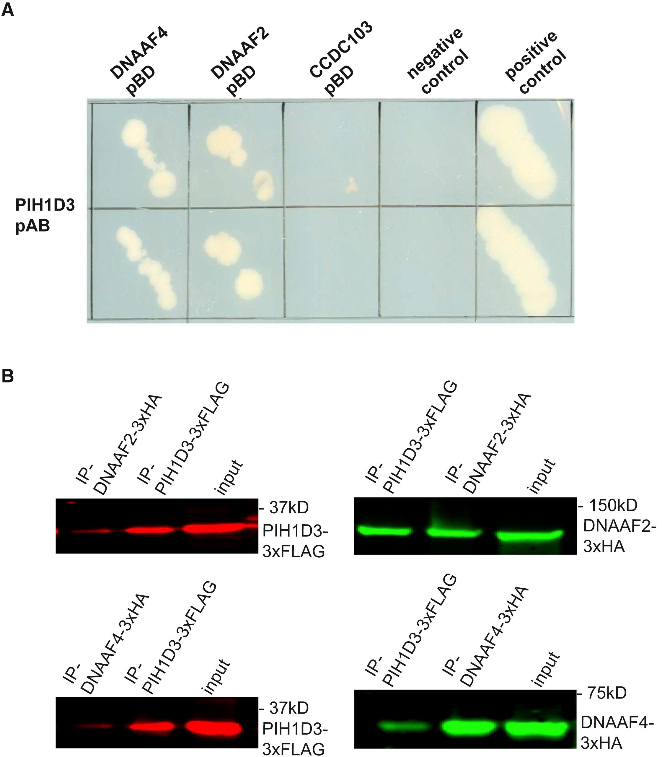
Figure 4: Identification of PIH1D3 Interacting Genes
(A) Y2H assays show strong interaction of PIH1D3 with DNAAF2 and DNAAF4. Lamin-C-pBD- Gal4-pAD was used as a negative control and Gal4-pBD - Gal4-pAD as a positive control.</br>(B) Confirmation of PIH1D3 interactors from the Y2H screen by co-IP from HEK293T cells. Co-expressions of tagged versions of PIH1D3 and either DNAAF2 or DNAAF4 revealed protein-protein interactions with DNAAF2 and DNAAF4 (left, western-blot anti-Flag; right, western blot anti-HA).
Licensed under: https://creativecommons.org/licenses/by-nc-nd/4.0/