Characterization of migratory primordial germ cells in the aorta-gonad-mesonephros of a 4.5-week-old human embryo: a toolbox to evaluate in vitro early gametogenesis
Maria GomesFernandes, Monika Bialecka, Daniela C F Salvatori, Susana M Chuva de Sousa Lopes, 08.03.2018
Abstract
STUDY QUESTIONWhich set of antibodies can be used to identify migratory and early post-migratory human primordial germ cells (hPGCs)? STUDY FINDING We validated the specificity of 33 antibodies for 31 markers, including POU5F1, NANOG, PRDM1 and TFAP2C as specific markers of hPGCs at 4.5 weeks of development of Carnegie stage (CS12–13), whereas KIT and SOX17 also marked the intra-aortic hematopoietic stem cell cluster in the aorta-gonad-mesonephros (AGM). WHAT IS KNOWN ALREADY The dynamics of gene expression during germ cell development in mice is well characterized and this knowledge has proved crucial to allow the development of protocols for the in vitro derivation of functional gametes. Although there is a great interest in generating human gametes in vitro, it is still unclear which markers are expressed during the early stages of hPGC development and many studies use markers described in mouse to benchmark differentiation of human PGC-like cells (hPGCLCs). Early post-implantation development differs significantly between mice and humans, and so some germ cells markers, including SOX2, SOX17, IFITM3 and ITGA6 may not identify mPGCs and hPGCs equally well. STUDY DESIGN, SIZE, DURATION This immunofluorescence study investigated the expression of putative hPGC markers in the caudal part of a single human embryo at 4.5 weeks of development. PARTICIPANTS/MATERIALS, SETTING, METHODS We have investigated by immunofluorescence the expression of a set of 33 antibodies for 31 markers, including pluripotency, germ cell, adhesion, migration, surface, mesenchymal and epigenetic markers on paraffin sections of the caudal part, including the AGM region, of a single human embryo (CS12–13). The human material used was anonymously donated with informed consent from elective abortions without medical indication. MAIN RESULTS AND THE ROLE OF CHANCE We observed germ cell specific expression of NANOG, TFAP2C and PRDM1 in POU5F1+ hPGCs in the AGM. The epigenetic markers H3K27me3 and 5mC were sufficient to distinguish hPGCs from the surrounding somatic cells. Some mPGC-markers were not detected in hPGCs, but marked other tissues; whereas other markers, such as ALPL, SOX17, KIT, TUBB3, ITGA6 marked both POU5F1+ hPGCs and other cells in the AGM. We used a combination of multiple markers, immunostaining different cellular compartments when feasible, to decrease the chance of misidentifying hPGCs. LARGE SCALE DATANon-applicable. LIMITATIONS REASONS FOR CAUTION Material to study early human development is unique and very rare thus restricting the sample size. We have used a combination of antibodies limited by the number of paraffin sections available. WIDER IMPLICATIONS OF THE FINDINGS Most of our knowledge on early gametogenesis has been obtained from model organisms such as mice and is extrapolated to humans. However, since there is a dedicated effort to produce human artificial gametes in vitro, it is of great importance to determine the expression and specificity of human-specific germ cell markers. We provide a systematic analysis of the expression of 31 different markers in paraffin sections of a CS12–13 embryo. Our results will help to set up a toolbox of markers to evaluate protocols to induce hPGCLCs in vitro. STUDY FUNDING AND COMPETING INTEREST(S) M.G.F. was funded by Fundação para a Ciência e Tecnologia (FCT) [SFRH/BD/78689/2011] and S.M.C.S.L. was funded by the Interuniversity Attraction Poles (IAP, P7/07) and the European Research Council Consolidator (ERC-CoG-725722-OVOGROWTH). The authors declare no conflict of interest.
GOMES FERNANDES, Maria, et al. Characterization of migratory primordial germ cells in the aorta-gonad-mesonephros of a 4.5-week-old human embryo: a toolbox to evaluate in vitro early gametogenesis. MHR: Basic science of reproductive medicine, 2018, 24. Jg., Nr. 5, S. 233-243.
Publication: https://doi.org/10.1093/molehr/gay011
 Disclaimer
The publication Characterization of migratory primordial germ cells in the aorta-gonad-mesonephros of a 4.5-week-old human embryo: a toolbox to evaluate in vitro early gametogenesis by Maria GomesFernandes, Monika Bialecka, Daniela C F Salvatori, Susana M Chuva de Sousa Lopes is published under an open access license: https://creativecommons.org/licenses/by-nc/4.0/. Permits non-commercial re-use, distribution, and reproduction in any medium, provided the original work is properly cited.
Curation by the MFGA team Relevant data sets presented in the publication have been identified. If possible, annotations (title, general information, conditions, processed tissue types and processed cell types) have been added based on information from the publication. Data tables and images that provide a good overview on the publication's findings on the data set have been extracted from the publication and/or supplement. If not stated otherwise, images are depicted with title and description exactly as in the publication. Tables have been adjusted to the MFGA table format. Conducted adjustments are explained in the detailed view of the tables. However, titles and descriptions have been adopted from the publication.
Data set 1: Evaluation of putative hPGC markers in the caudal part of a single human embryo at 4.5 weeks of development
Other: Immunohistochemistry
Species
| Species |
|---|
| Human |
Tissue Types
| BRENDA tissue ontology | Maturity | Description | Species | Replicates |
|---|---|---|---|---|
| BTO_0000534: gonad | Embryonic | W 8-9 | Human | 1 |
| Embryonic | W 9 | Human | 1 | |
| BTO_0000671: kidney | Embryonic | W 16-18 | Human | 1 |
| Embryonic | W 19 | Human | 1 | |
| BTO_0001078: placenta | Embryonic | W 19 | Human | 1 |
| BTO_0000269: colon | Embryonic | W 15 | Human | 1 |
Cell Types
| Cell ontology | Maturity | Description | Species | Replicates | Cells per replicate |
|---|---|---|---|---|---|
| CL_0000670: primordial germ cell | Primordial | hPGCs | Human | 1 | |
| Primordial | hPGCLCs | Human | 1 |
Data set 2: Early hPGCs showed a distinct epigenetic state from the somatic compartment
Methylome: Other
Species
| Species |
|---|
| Human |
Tissue Types
| BRENDA tissue ontology | Maturity | Description | Species | Replicates |
|---|---|---|---|---|
| Embryonic | Aorta-gonad-mesonephros (AGM) of CS12–13 human embryo | Human | 1 |
Cell Types
| Cell ontology | Maturity | Description | Species | Replicates | Cells per replicate |
|---|---|---|---|---|---|
| CL_0000670: primordial germ cell | Primordial | Human primordial germ cells (hPGC) | Human | 1 | |
| CL_0002371: somatic cell | Embryonic | Human | 1 |
Images
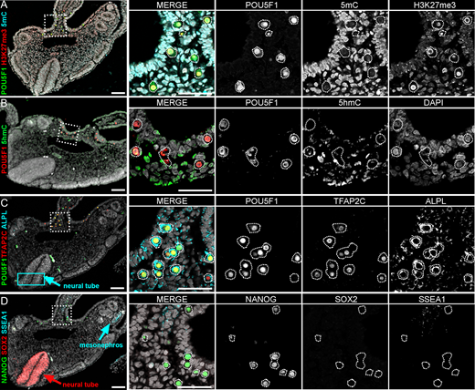
Figure 2: Expression of pluripotency markers in the AGM of a CS12–13 human embryo
A-D: Histological sections of the caudal part of the embryo immunostained for POU5F1 (green), H3K27me3 (red) and 5-methylcytosine (5mC, cyan) (A); POU5F1 (red) and 5-hydroximethylcytosine (5hmC, green) (B); POU5F1 (green), TFAP2C (red) and ALPL (cyan; cyan box depicts staining in neural tube) (C); and NANOG (green), SOX2 (red) and SSEA1 (cyan) (D). All sections were counterstained with DAPI (grey). Right panels depict a higher magnification corresponding to the dashed box in the left column (merge and single channels). Scale bars are 100 μm in the left column and 50 μm in all high magnifications.
Licensed under: https://creativecommons.org/licenses/by-nc/4.0/
Data set 3: Expression of mPGC-markers, mesenchymal and adhesion molecules in early hPGCs and signalling pathways
Other: Immunohistochemistry
Species
| Species |
|---|
| Human |
Tissue Types
| BRENDA tissue ontology | Maturity | Description | Species | Replicates |
|---|---|---|---|---|
| Embryonic | aorta-gonad-mesonephros of CS12–13 human embryo | Human | 1 | |
| BTO_0000315: ectoderm | Embryonic | Surface Ectoderm | Human | 1 |
| BTO_0001768: notochord | Embryonic | Human | 1 | |
| BTO_0001057: neural tube | Embryonic | Human | 1 | |
| BTO_0000545: gut | Embryonic | Human | 1 | |
| BTO_0000742: myotome | Embryonic | Human | 1 | |
| BTO_0006248: dermatome | Embryonic | Human | 1 | |
| BTO_0002045: capillary | Embryonic | Human | 1 |
Cell Types
| Cell ontology | Maturity | Description | Species | Replicates | Cells per replicate |
|---|---|---|---|---|---|
| CL_0000670: primordial germ cell | Primordial | Human primordial germ cells (hPGCs) | Human | 1.0 | |
| CL_0000115: endothelial cell | Embryonic | Human | 1.0 | ||
| CL_0002371: somatic cell | Embryonic | Human |
Images
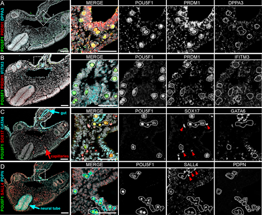
Figure 3: Expression of germ cell-associated markers in the AGM of a CS12–13 human embryo
A-D: Histological sections of the caudal part of the embryo immunostained for POU5F1 (green), PRDM1 (red) and DPPA3 (cyan) (A); POU5F1 (green), PRDM1 (red) and IFITM3 (cyan) (B); POU5F1 (green), SOX17 (red) and GATA6 (cyan) (C); and POU5F1 (green), SALL4 (red) and PDPN (cyan) (D). All sections were counterstained with DAPI (grey). Right panels depict a higher magnification corresponding to the dashed box in the left column (merge and single channels). Scale bars are 100 μm in the left column and 50 μm in all high magnifications.
Licensed under: https://creativecommons.org/licenses/by-nc/4.0/
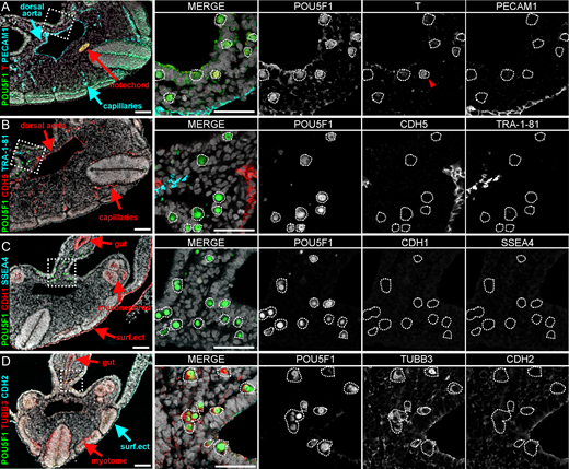
Figure 4: Expression of mesenchymal and adhesion molecules in the AGM of a CS12–13 human embryo
A-D:Histological sections of the caudal part of the embryo immunostained for POU5F1 (green), T (red) and PECAM1 (cyan) (A); POU5F1 (green), CDH5 (red) and TRA-1–81 (cyan) (B); POU5F1 (green), CDH1 (red) and SSEA4 (cyan) (C); and POU5F1 (green), TUBB3 (red) and CDH2 (cyan) (D). All sections were counterstained with DAPI (grey). Right panels depict a higher magnification corresponding to the dashed box in the left column (merge and single channels). Scale bars are 100 μm in the left column and 50 μm in all high magnifications. surf.ect, surface ectoderm.
Licensed under: https://creativecommons.org/licenses/by-nc/4.0/
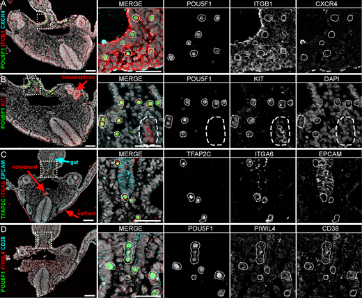
Figure 5: Expression of migratory and surface markers in the AGM of a CS12–13 human embryo
A-D: Histological sections of the caudal part of the embryo immunostained for POU5F1 (green), ITGB1 (red) and CRCXR4 (cyan) (A); POU5F1 (green), KIT (red) and DAPI (grey) (B); TFAP2C (green), ITGA6 (red) and EPCAM (cyan) (C); and POU5F1 (green), PIWIL4 (red) and CD38 (cyan) (D). All sections were counterstained with DAPI (grey). Right panels depict a higher magnification corresponding to the dashed box in the left column, as merge and each single channel except DAPI. Scale bars are 100 μm in the left column and 50 μm in all high magnifications. surf.ect, surface ectoderm.
Licensed under: https://creativecommons.org/licenses/by-nc/4.0/